News Story
Aristos Christou Heads Project for GaN Amplifiers in MRI Systems
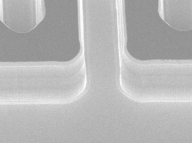
Image: Deep reactively trenched in GaN required for new Trench GaN MOSFET - the trench is 5 microns deep.
Aristos Christou, a materials science and engineering professor at the University of Maryland (UMD), recently began a new research project on research in the area of gallium nitride (GaN) based power semiconductor switches for ultra-high field magnetic resonance imaging, or MRI systems. The study, entitled, "Optimization of Power Semiconductor Switch for RF On-Coil Amplification for Ultra-High Field MRI," will be conducted in collaboration and leadership with the National Institutes of Health (NIH) scientists, Drs. Natalia Gudino and Jeff Duyn.
"Most high frequency power amplifiers used in current MRI systems are built either with metal oxide semiconductor (MOS) field effect transistor (FET) or with metal semiconductor transistors (MESFETs)," Christou said. "GaN and silicon carbide (SiC) are excellent materials for building high-power/high-frequency switching devices for use at high voltages and high magnetic fields. The reason for this is due to their wide band gap, high breakdown voltage and high mobility of electrons in a conducting channel. The high electron mobility can be further increased if the conducting channel comprises of a sheet of electrons so that a nearly two dimensional sheet is attained. These devices, called high electron mobility transistors (HEMTs), can be combined into a complex circuit, which helps reduce parasitic components and manufacturing cost. However, current devices fail to meet the requirements for an optimum power amplifier, capable of operating efficiently in the ultra-high magnetic field MRI environment."
To that end, Christou and his team set about engineering a device for this specific application.
The researchers aim to assess the performance of commercially available FETs in the magnetic resonance environment. Previous studies suggest that the performance of such devices, normally located on or near the MRI coil, can degrade with increasing magnetic field. Since most MRI power amplifiers are located outside the MRI room, there is a lack of data on the performance and reliability of GaN based FETs when they are immersed in the MR environment. Recognizing these limitations, NIH scientists sought to collaborate with Christou in order to understand the degradation phenomenon, and then design and fabricate optimized FETs capable of withstanding the MR environment.
NIH has been a leader in advanced MRI systems. The Nuclear Magnetic Resonance (NMR) Center at NIH has MRI systems with a wide range of magnetic field strengths, which provides a perfect site for analyzing the effects of the magnetic field on various device properties, especially those relevant to achieving high performance of the on-coil power amplifier. Moreover, Christou and materials science graduate student, Brett Setera, will investigate GaN based FET switches which will be fabricated in the clean room of the UMD Nanocenter.
"With the development of these GaN amplifiers, we expect to enable a new MRI technology, and scientists will be able to precisely image artifacts in the brain, with enhance resolution and sensitivity, which have remained elusive to medical doctors," said Christou.
Published July 8, 2021
Recent Stories
Stories / May 26, 2026
Apply: Chair of the Fischell Dept of Bioengineering

Stories / May 28, 2026
Generations of Graduates: A Full-Circle Celebration

Stories / May 28, 2026
Student Spotlight: Outstanding Junior Award Recipient Joel...

Stories / May 28, 2026
New Framework Could Speed Up Thermal Storage Design and Reduce...

Stories / May 28, 2026
UMD Alum Wins ACM SIGMOBILE Dissertation Award

Stories / May 26, 2026
ECE Faculty and Staff Honored by The Universities of Shady Grove

Stories / May 26, 2026
Air Quality Study on 2023 Canadian Wildfires Reports New...

Stories / May 22, 2026
ECE PhD Student Sydney Overton Receives Ann G. Wylie...

Stories / May 21, 2026
SMILE: Automated Swing Delivers Real-World Impact for Maryland...

Stories / May 20, 2026
UMD Research to Study Aerosol Transmission of Respiratory...
