News Story
Scarcelli Applies NSF CAREER Award to Study Embryonic Development
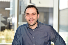
Dr. Giuliano Scarcelli
Fischell Department of Bioengineering (BIOE) faculty member Giuliano Scarcelli was named a recipient of the National Science Foundation’s Faculty Early Career Development (CAREER) award. The five-year, $537,000 award will support Scarcelli’s efforts to develop a novel imaging technology to measure how biological cells grow into complex tissues during embryonic development.
Currently a BIOE assistant professor, Scarcelli was recently promoted to associate professor, effective July 1. He is also a faculty member of the University of Maryland’s Robert E.Fischell Institute for Biomedical Devices.
Applying a technique known as Brillouin microscopy, Scarcelli and members of his Optics Biotech Lab are working to improve understanding of the biomechanical aspects of embryonic development. Previous technologies to observe embryonic development have mostly focused on probing genetic and biochemical control of tissue growth.
“While our understanding of genetic, biochemical, and molecular controls of tissue growth can rely on several established methods, the biomechanics of tissue morphogenesis remains poorly understood mostly due to the lack of suitable measurement techniques,” Scarcelli said.
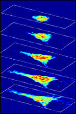
Brillouin microscopy relies on a specialized light-scattering phenomenon to image the viscoelastic and mechanical properties of biological materials in a non-invasive manner. In this project, Scarcelli and his team are taking the technology a step further by developing a form of “light-sheet” Brillouin microscopy, such as where all points illuminated by a thin optical beam are measured at once.This will enable rapid mapping of elastic modulus in a developing embryo — without contact — at high 3D resolution.
“Brillouin microscopy has already proven impactful in tissue and cell biomechanics, but it currently relies on point-scanning to build an image,” Scarcelli said. “Thus, traditional Brillouin microscopy techniques are not suited for embryo studies due to speed and damage limitations.”
By developing a Brillouin light-scattering technology compatible with light-sheet microscopy platforms, Scarcelli and his team will be able to superimpose their mechanical maps with structural and functional properties of tissues and cells. This, in turn, enables the group to advance mechanobiology studies of morphogenesis by allowing them to observe properties such as elasticity, as well as cell and tissue size, shape, and organization, in addition to cell signaling activation and expression patterns.
Working at the interface of photonics, biomechanics and developmental biology, Scarcelli and his team plan to use NSF support to provide opportunities to educate and train graduate and undergraduate students with a diverse set of skills. Through the project, Scarcelli also hopes to introduce middle-school students to STEM through activities that offer hands-on exercises that connect microscopy technology to real-life familiar applications.
Published June 15, 2020
Related Stories
Stories / September 16, 2024
Dutt Receives NSF CAREER Award

Stories / January 18, 2024
Dutta to receive NSF CAREER Award

Stories / June 16, 2023
Erika Moore Receives NSF CAREER Award
Stories / May 15, 2023
UMD’s Tubaldi Wins NSF CAREER Award

Stories / February 2, 2022
Allison Reilly Wins NSF CAREER Award

Stories / January 27, 2022
Daniels Receives NSF CAREER Award

Stories / December 3, 2021
You Zhou Receives NSF CAREER Award

Stories / May 14, 2021
Four Clark School Faculty Receive CAREER Awards

Stories / February 2, 2021
Maisel Applies NSF CAREER Award to Advance Drug Delivery...
Stories / October 5, 2015
Novel Microscopy Technique to Shed New Light on Study of Cell...