News Story
Uncovering the mysteries of networking in the brain
What’s going on with the neurons inside our brains when we gather information through our senses, interpret what it means, and act on our decisions? New work by University of Maryland researchers and their colleagues published in the journal Cell Reports is providing some answers.
When mammals navigate their environment, their brains must process both sensory information and context so that they can behave appropriately. But scientists do not yet know how individual neurons and the cortical networks they form implement this information processing.
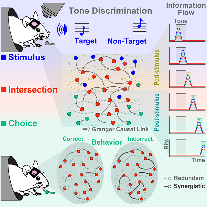
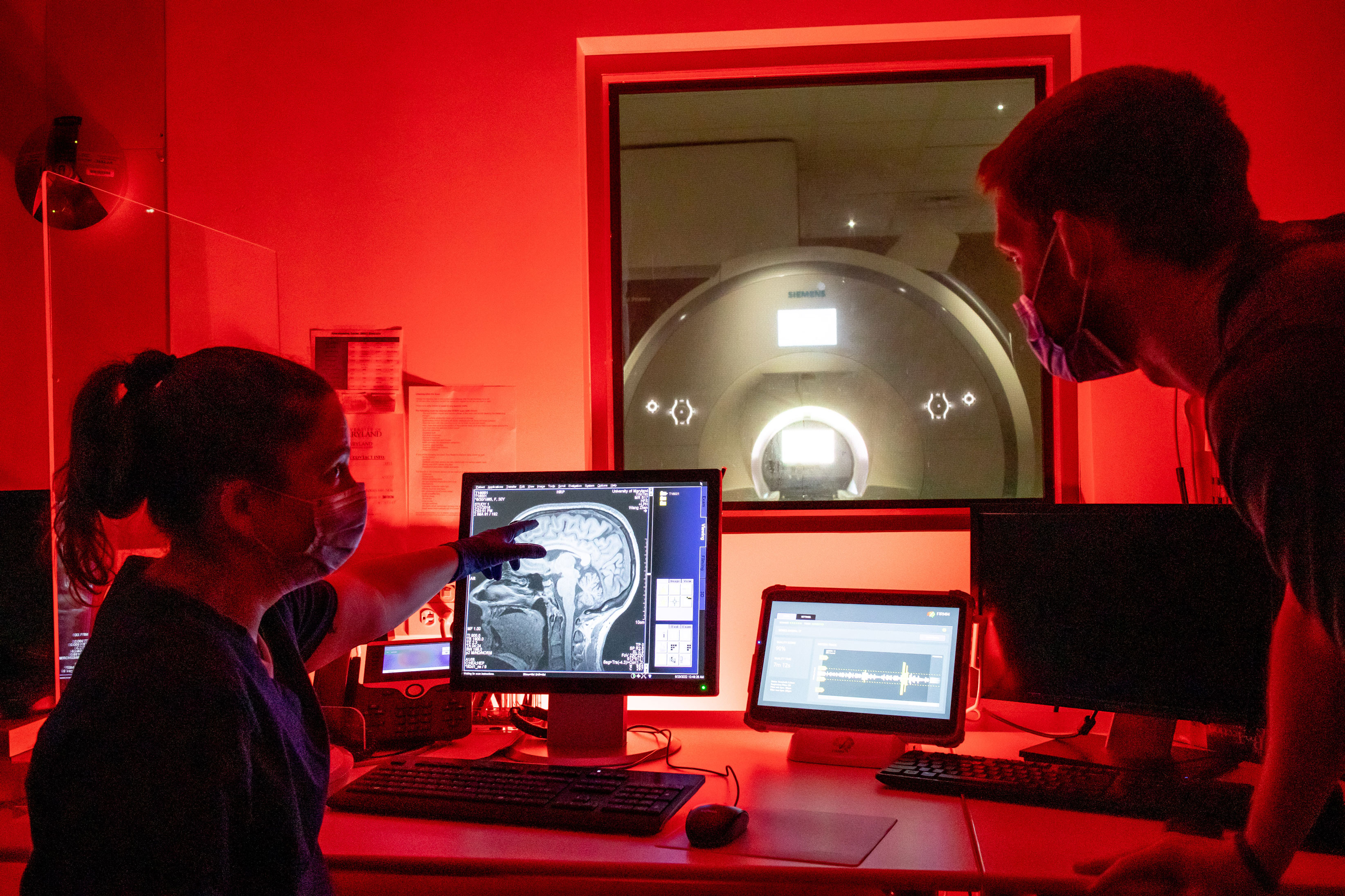
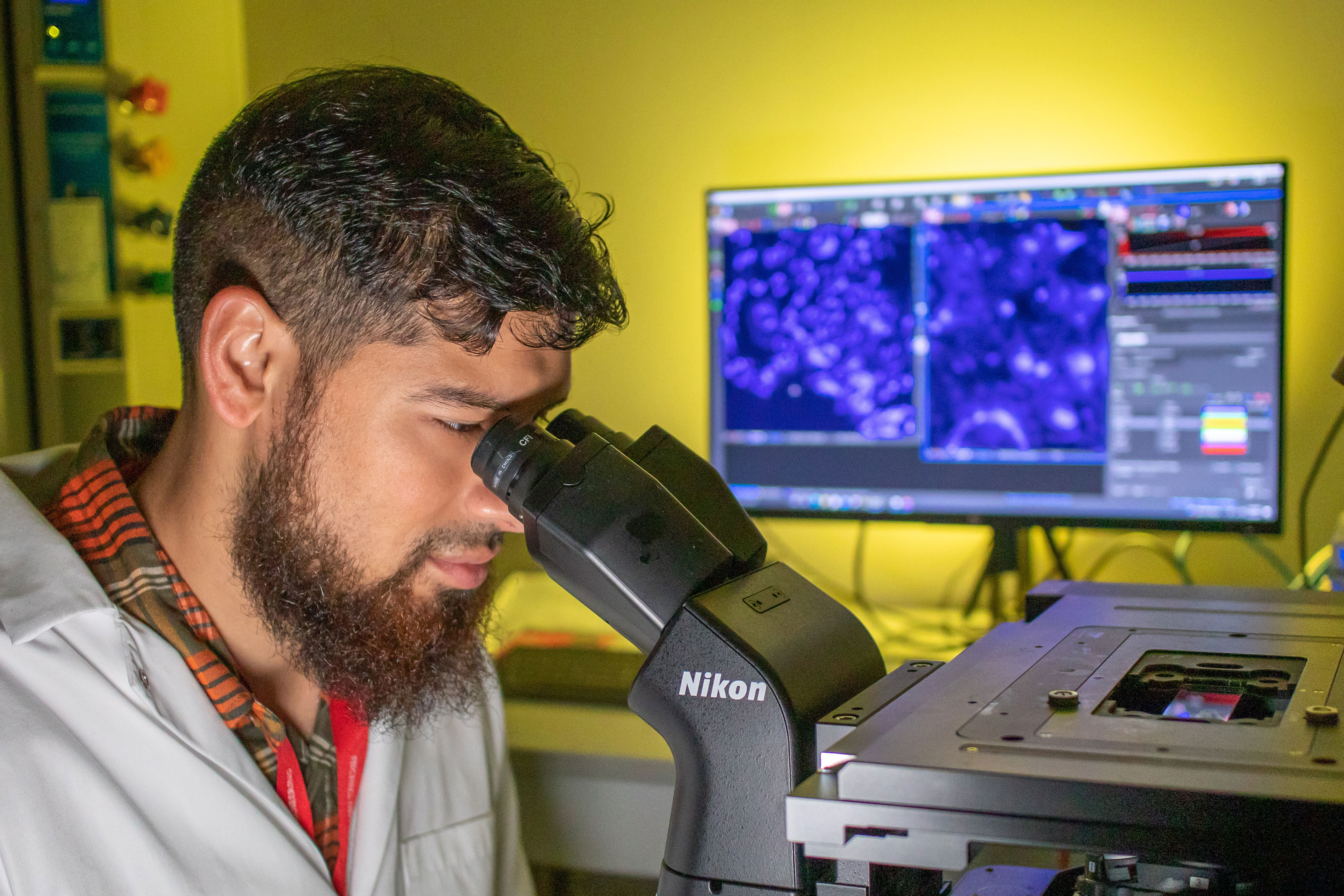
In “Sequential transmission of task-relevant information in cortical neuronal networks,” the researchers used two-photon imaging to record neuronal activity from the primary auditory cortex of mice during a pure-tone discrimination task, which requires mice not only to recognize which pure-tone is being presented but its meaning as well (i.e. that the “target” tone is rewarded while the “non-target” tone is not). Their findings describe specialized neurons that integrate task-related information and form functional networks whose structures encode both sensory input and behavioral choice.
The researchers found that while some neurons carry only acoustic information and some carry information about the choice the animal has made, others specifically carry the intersection of the two – i.e., information about sensory input used to inform the animal’s choice. Further examination of these “intersection neurons” showed that although individually they carry information transiently, they formed networks in which task-relevant information persisted and was sequentially transmitted between neurons throughout the duration of a task.
In addition, the information content of intersection neurons changed depending on a mouse’s decision. When a mouse made a correct decision, the information shared between functionally connected intersection neurons was mostly redundant—neurons carried similar information. However, when the animal made a mistake, the shared information was synergistic, meaning that the neurons carried distinct information.
“Most existing work considers encoding of the external stimuli as a separate process from the readout of information in the brain that results in behavior,” said Associate Professor Behtash Babadi (ECE/ISR), one of the paper’s authors. “Our results show that there are indeed specialized groups of neurons in the auditory cortex that integrate the two processes by carrying both the information about the external stimuli and the animal’s behavior sequentially through time. The manner by which they carry information matters for correct vs. incorrect behavior.”
The paper was published in Cell Reports on May 31. This is collaborative work by the labs of Professor Patrick Kanold (formerly UMD, now Biomedical Engineering, Johns Hopkins University), Associate Professor Behtash Babadi (ECE/ISR), and Dr. Stefano Panzeri (Laboratory of Neural Computation, Istituto Italiano di Tecnologia in Italy and the Center for Molecular Neurobiology, University Medical Center, Hamburg-Eppendorf, Germany). Co-lead authors are Kanold’s former postdoctoral fellow and current Assistant Professor Nikolas Francis (UMD Biology/BBI), Babadi’s Ph.D. student Shoutik Mukherjee (UMD ECE), and Panzeri’s postdoctoral fellow Loren Koçillari.
The research is funded by a National Institutes of Health “U19” grant awarded in 2019. “Readout and control of spatiotemporal neuronal codes for behavior” is a five-year, $20M project in NIH’s BRAIN Initiative.
This large project is providing a clearer picture of how the brain operates in real time, with sensory input, brain activity, and behavior interacting in complex, simultaneous, mutually informing ways. It is revealing how the external world shapes the patterns of brain activity, brain activity manifests in behavior, and the elicited behavior in turn reshapes the brain.
This paper is a part of “Determining which Neurons Contribute to a Particular Behaviorally Distinguishable Percept,” a project within the main grant that has the goal of establishing causal links between neural activity and behavior.
“An exciting consequence of these results is that we can now target these ‘intersection neurons’ using holographic optogenetics, and test whether the animal’s behavior can be altered by manipulating the network structure of these neurons” says Babadi. “Ultimately, a clear understanding of how neural activity and behavior affect one another can positively impact neural prostheses and brain-machine interface systems.”
U19s are among the largest awards given by NIH, and are dedicated to highly ambitious, multi-disciplinary, multi-university projects. The overall project is led by John H.R. Maunsell, Albert D. Lasker Distinguished Service Professor of Neurobiology and Director of the Grossman Institute for Neuroscience at the University of Chicago. The subproject featuring UMD researchers is led by Patrick Kanold.
Published June 14, 2022
Related Stories
Stories / October 25, 2021
How does the brain turn heard sounds into comprehensible...
Stories / April 28, 2023
Autism Research Resonates in Hearing-Focused Project
Stories / September 9, 2022
$7.9 Million in NIH Awards Propel UMD Aging Research
Stories / November 1, 2021
How Home Alone Helped UMD Neuroscientists Unlock Brain Scan Data
Stories / September 9, 2020
Cornelia Fermüller is PI for 'NeuroPacNet,' a $1.75M NSF...

Stories / December 7, 2023
‘Priming’ helps the brain understand language even with...
Stories / September 26, 2023
New UMD Division of Research video highlights work of Simon,...

Stories / September 30, 2022
Do Suddenly Self-Centered Brain Cells Promote Disease?
Stories / September 2, 2022
New robust and scalable computational methodology developed by...
Stories / December 20, 2021
2022 BBI Seed Grant Awards to Fuel Innovation in Aging and...