News Story
UMD research team creates ‘switchable’ adhesive for repairing cuts and tears in tissue
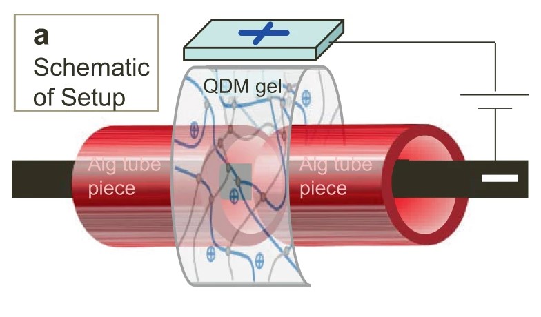
A long QDM gel-strip is used as a sleeve around two pieces of the Alg tube.
Polymers, specifically hydrogels, have become a household staple in modern society. Examples include disposable contacts and Jell-O, although gels can also be found in hair products, toothpaste, various cosmetics and even disposable diapers. Hydrogels are soft, flexible and boast high water content due to their hydrophilic nature, which is similar to human tissue, making the material a good candidate for medical applications.
Srinivasa R. Raghavan, a chemical and biomolecular engineering (ChBE) professor at the University of Maryland (UMD), and his research team frequently experiment with soft, responsive nanomaterials, including hydrogels. Leah Borden, a ChBE Ph.D. Student working with Dr. Raghavan, has always been fascinated by the medical field and was keen to analyze a problem that could have a positive impact on human health.
“I tend to look at problems through a medical lens, so when the idea to adhere gels to tissue was suggested by Dr. Raghavan, I started looking at tubes in the body that might need repair, and blood vessels made the most sense,” said Borden, who served as first author on the study, published in Nature Communications. “I purchased bovine tissue from a local butcher shop. Specifically, I cut a piece of the aorta (the main artery) and sandwiched it together with a non-sticky hydrogel. Then, I applied an electric current (10 volts of DC for 20 seconds) to the 'gel-aorta sandwich.' To my surprise, the two materials stuck together strongly. When I reversed the polarity of the current, the gel could be easily removed from the tissue without causing any damage.”
Borden realized that she had discovered a new way to switch the adhesion between the gel and the tissue on and off, calling this phenomenon "electroadhesion." The researchers found that electroadhered gel-patches can provide a seal over cuts, or openings, in bovine aorta, or other tissues, while a gel “sleeve” could rejoin pieces of a severed tube. Electroadhesion works with many, but not all tissues and the team is trying to figure out why.
The potential for this technique in surgery, however, is clear – it could replace the need for expensive, time-consuming sutures, which need to be applied by a medical professional. It could also replace unsuccessful practices such as the use of surgical glues (which adhere weakly), or failed repairs by techniques such as catherization.
"We’ve had significant interest in the surgical applications of electroadhesion!" Borden said. "We're collaborating with surgeons at the University of Chicago and Children’s National in DC, among others, and we hope to begin animal trials in the near future."
This process could also have applications in soft robotics, 3D printing, the automotive industry, and beyond.
The team also authored a blog-post for the Nature Bioengineering Community website, describing the potential applications of this technology.
For additional information:
Borden, L.K., Gargava, A. & Raghavan, S.R. (2021). Reversible electroadhesion of hydrogels to animal tissues for suture-less repair of cuts or tears. Nat Commun 12, 4419. DOI: 10.1038/s41467-021-24022-x
Raghavan, S.R. A new discovery that could revolutionize surgery: Gels can be adhered to animal tissues by applying an electric field.
Published September 22, 2021
Recent Stories
Stories / May 26, 2026
Apply: Chair of the Fischell Dept of Bioengineering

Stories / May 28, 2026
Student Spotlight: Outstanding Junior Award Recipient Joel...

Stories / May 26, 2026
ECE Faculty and Staff Honored by The Universities of Shady Grove

Stories / May 26, 2026
Air Quality Study on 2023 Canadian Wildfires Reports New...

Stories / May 22, 2026
ECE PhD Student Sydney Overton Receives Ann G. Wylie...

Stories / May 21, 2026
SMILE: Automated Swing Delivers Real-World Impact for Maryland...

Stories / May 20, 2026
UMD Research to Study Aerosol Transmission of Respiratory...

Stories / May 19, 2026
Ambi Narula Honored with Pride and Advocacy Award

Stories / May 19, 2026
Student Success at SAMPE 2026

Stories / May 19, 2026
New UMD Patent Advances Compact, High-Performance Phase Change...
