News Story
Research Team Uses Dark Field Optical Microscopy to 'See' Nanoparticles
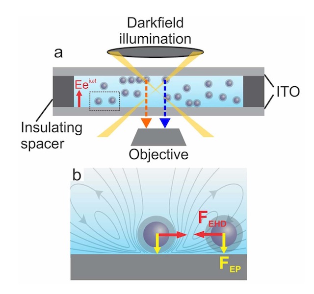
Image (not to scale): Schematic of sample cell and dark field optical microscopy for visualizing AC electric field induced NP assembly.
Researchers in the Department of Chemical and Biomolecular Engineering (ChBE) at the University of Maryland (UMD) have developed a new method for using optical microscopy to observe how nanoparticles behave and self-assemble in electric fields.
Arranging nanoparticles into hierarchically structured materials – displaying order on different length scales – is a challenge of nanoscience research due to the fact that these materials have desirable functional properties (e.g., optical, electrical, or mechanical) far exceeding those that exist in bulk materials. For example, assemblies of precious metal nanoparticles, like gold or silver nanoparticles, have optical properties allowing them to act as biosensors, or advanced optical coatings (device displays, for example) – carefully organizing these nanoparticles would greatly enhance efficiency and selectivity.
Scientists have tried to create different ways to form hierarchically structured materials. One current method of assembling nanoparticles is to dissolve them in a liquid phase and apply external electric fields as a means of ‘directing’ the nanoparticles into a desired configuration – a complex and ambiguous process, due in large part to the fact that no one has ever directly seen how the nanoparticles behave when an electric field is applied – nanoparticles are too small to be seen with conventional microscopy. Without direct observation, it is impossible to develop physics models for how the nanoparticles will behave in electric fields under various conditions.
To that end, ChBE Assistant Professor Taylor Woehl and his research group, have developed an advanced type of optical microscopy, called dark field optical microscopy, in conjunction with a special type of nanoparticles, called plasmonic nanoparticles. Adam Ferrick - a ChBE undergraduate student who is graduating this spring - served as first author on the study, published May 4, 2018, in Langmuir.
“In this study, we use plasmonic silver nanoparticles, which due to their plasmonic properties, strongly scatter blue light,” said Dr. Woehl. “This allows us to indirectly ‘see’ the nanoparticles by taking microscopy images of the blue light that they emit. When the silver nanoparticles come close to each other during assembly, the light they scatter becomes more red in color due to a phenomenon called interparticle plasmonic coupling. So, even though we can’t see the individual nanoparticles as they come close to each other, we indirectly know this is happening by observing a color change in our microscopy images.”
Basically the nanoparticles sense each other when they get close enough together and start to behave as a single, larger nanoparticle.
The group has used this approach to investigate how nanoparticles behave in alternating-current electric fields. By imaging color changes with dark field microscopy, they found nanoparticles form large 2-dimensional assemblies on the electrode surface.
“We have developed a physics model to describe this behavior and find that the assembly is due to a phenomenon called electrohydrodynamic fluid flow,” said Woehl. “This specific process has actually been a mystery in the field for many decades – our experiments are the first visual confirmation of the what causes the formation of these 2-dimensional assemblies.”
In terms of contemporary applications, this system could take advantage of color change that occurs when an electric field is applied to create ‘active’ camouflage, or materials that change color on demand – almost like a synthetic version of octopus skin.
For additional information:
Adam Ferrick, Mei Wang and Taylor J. Woehl. “Direct Visualization of Planar Assembly of Plasmonic Nanoparticles Adjacent to Electrodes in Oscillatory Electric Fields.” Langmuir, 04 May 2018. DOI: 10.1021/acs.langmuir.8b00992
Published May 15, 2018
Related Stories
Stories / November 20, 2019
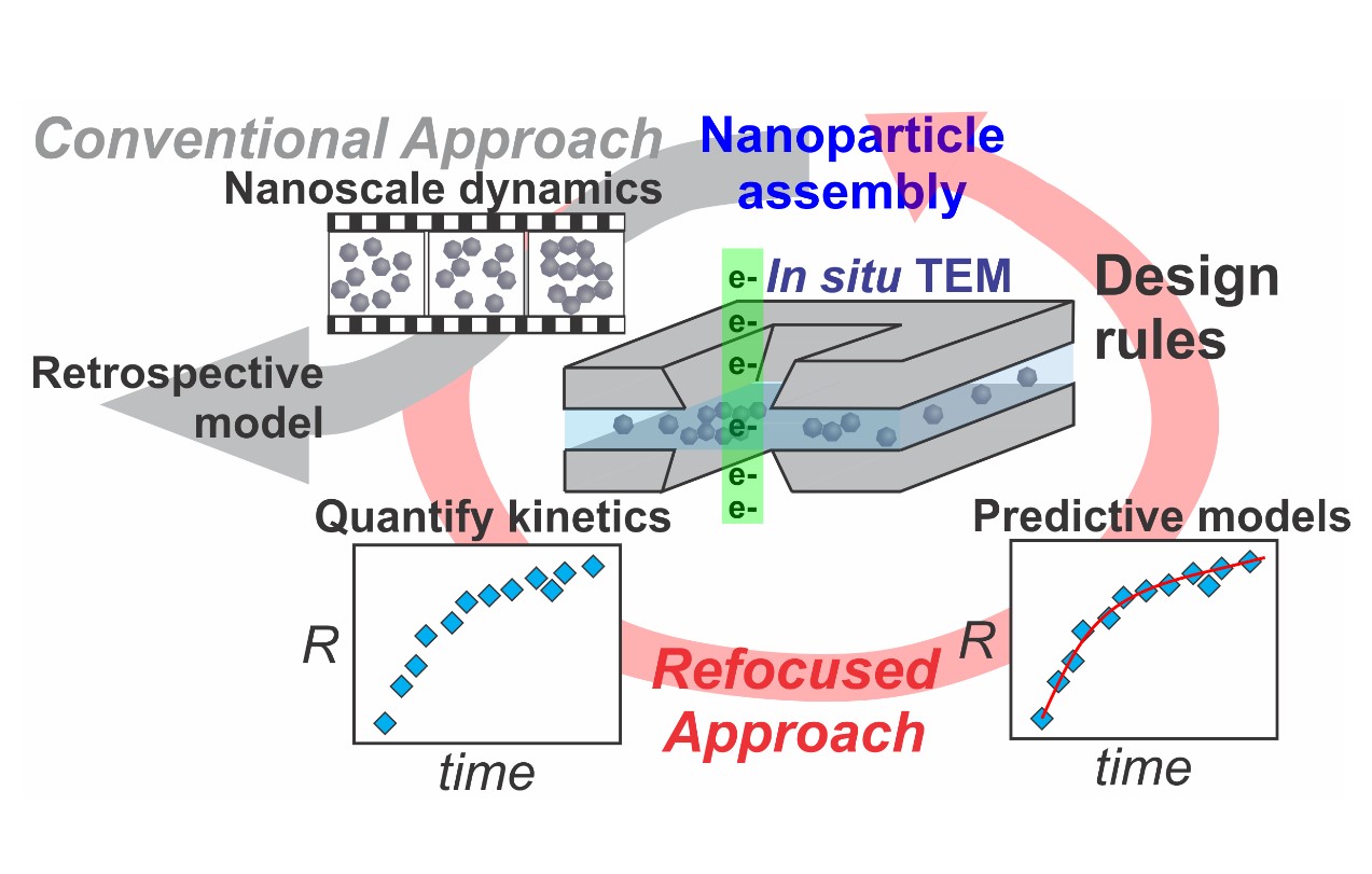
Refocusing In-situ Electron Microscopy
Stories / August 20, 2019
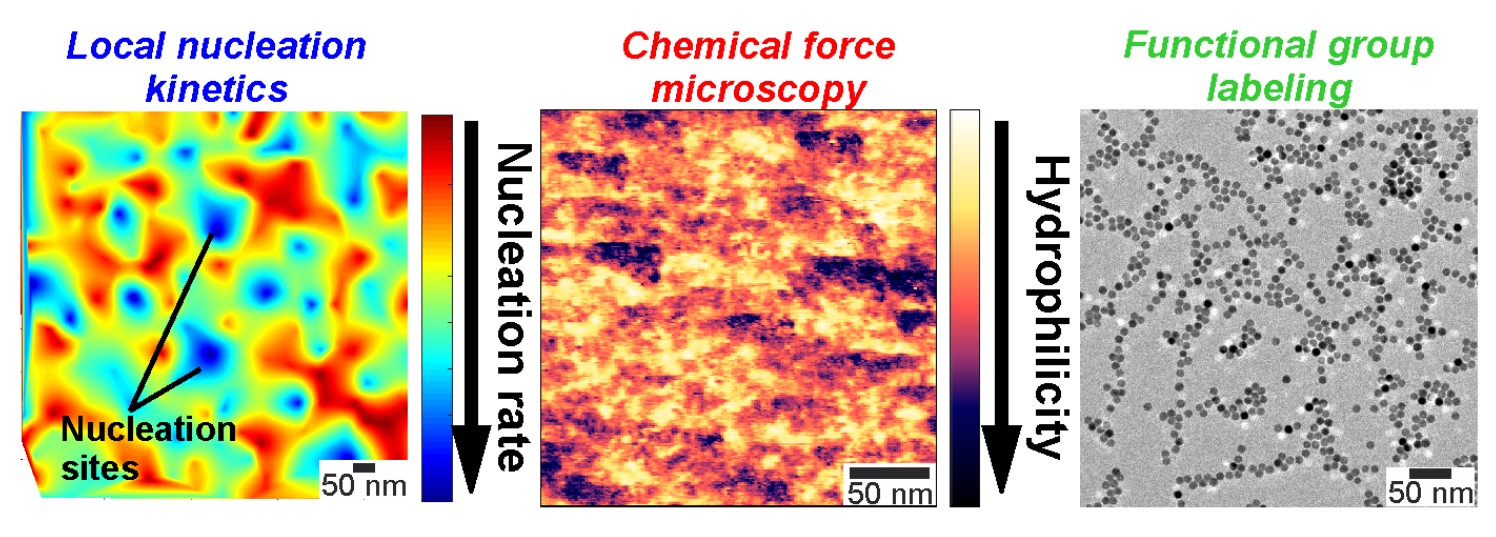
Mapping Nucleation Kinetics with Nanometer Resolution
Stories / Mar 13, 2026
Endless Possibilities for Life-Changing Good

Stories / Mar 12, 2026
Tim Koeth Named Exemplary Honoree at Maryland Research...

Stories / Mar 10, 2026
Wereley Named Fellow of the Canadian Society for Mechanical...

Stories / Mar 10, 2026
ION Storage Systems Announced Successful Customer Qualification

Stories / Mar 9, 2026
Engineering safer, more sustainable AI for all

Stories / Mar 6, 2026
New Faculty Spotlight: Netanel Korin

Stories / Mar 4, 2026
Wachsman comments on solid-state battery readiness

Stories / Mar 3, 2026
At Engineering’s Alumni Cup, the Atmosphere Was Electric

Stories / Mar 2, 2026
Tamunobelema Olungwe Gives Back

Stories / Mar 2, 2026
A New Chapter for Chelsea Neumann
