News Story
New Brain Probe Could Help Surgeons Avoid Blood Vessel Damage
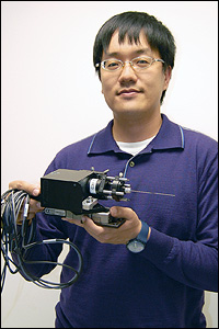
BioE graduate student Chia-Pin Liang holding the prototype Doppler Optical Coherence Tomography (DOCT) probe, which is designed to help surgeons navigate the brain and avoid damage to its blood vessels. Liang says that with further development, a handheld version could be created for other kinds of procedures in which doctors must guide tools deep into the body.
A new bioimaging probe developed at the Clark School for use in neurosurgery can produce detailed, real-time images from deep inside the brain. It is designed to help doctors detect and avoid blood vessels that can accidentally be damaged during a procedure.
Fischell Department of Bioengineering (BioE) graduate student Chia-Pin Liang, advised by BioE assistant professor Yu Chen, was presented with a 2012 Optical Coherence Tomography News Student Travel Grant Award for his presentation of the device.
In stereotactic neurosurgery, needle-based instruments are inserted into the brain and guided to coordinates calculated before the operation through the use of magnetic resonance imaging (MRI) or a computed tomography (CT) scan. These procedures are risky, because the tools are likely to lacerate blood vessels in their path, and because the leakage of cerebrospinal fluid during surgery can shift the brain, moving the target from its pre-determined location. Cerebral hemorrhages, stroke, and the death of the patient are all possible outcomes of these otherwise life-saving procedures.
To help reduce these risks, Liang and his colleagues, including doctors from the University of Maryland School of Medicine's (UM-SOM) Departments of Neurology and Neurosurgery and the Baltimore VA Medical Center (BVAMC), designed a needle-like probe that can travel with the surgical tools in a tube called a cannula, looking ahead and interpreting the biological landscape for the surgeon, who can then make course corrections that avoid blood vessels.
The device is based on Optical Coherence Tomography (OCT) technology, which produces micron-scale imaging of tissue in the body and in real time. It enables what Chen refers to as an "optical biopsy"—visualization of changes to tissues without the need for a minor surgery to acquire a sample. OCT is similar in concept to ultrasound, but creates images by measuring the echo time delay and intensity of back-reflected light rather than sound.
In addition to forward imaging, the probe was designed with an outside diameter of less than 1 mm to allow integration with existing surgical tools, Doppler imaging technology, and a high imaging rate to provide real-time feedback to the surgeon. The device's relatively low manufacturing cost creates the potential for a disposable version.
During tests, the prototype device was able to detect and quantify blood flow, as well as differentiate between arteries and veins. Tests on extracted human brain tissue demonstrated that the position of the probe's tip could be determined from the micro-anatomical landmarks it saw and relayed back to the user.
The next phase of development will be the miniaturization of the external portion of the device.
"We plan to construct a pen-sized imaging device," says Liang. "A robust handheld probe will further broaden its medical application—for example, it could help doctors find arteries during cardiac catheterization procedures."
Liang plans to use his award to attend the Optical Society's Biomdical Optics Conference, to be held in Miami in April.
Liang's work on the Doppler OCT imaging needle for neurosurgical application was originally funded by a University of Maryland-Baltimore/University of Maryland, College Park, seed grant. His collaborators on the project include Chen, Jeremiah Wierwille (BioE, Ph.D. '11), Wei Gong (BioE), Thais Moreira (BVAMC), Gary Schwartzbauer (UM-SOM), M. Samir Jafri (UM-SOM), and Chia-Min Tang (UM-SOM/BVAMC).
Published January 24, 2012
Recent Stories
Stories / May 26, 2026
Apply: Chair of the Fischell Dept of Bioengineering

Stories / Jun 9, 2026
The Future of Terps Racing is Electric

Stories / Jun 8, 2026
Alchemity receives Build our Future Award from Governor Moore

Stories / Jun 8, 2026
Shuna Ni Receives NSF CAREER Award

Stories / Jun 5, 2026
UMD Senior Joshua Mathew Wins 2026 Astronaut Scholarship

Stories / Jun 4, 2026
BIOE Graduate Program Receives Mentoring Excellence Award

Stories / Jun 4, 2026
Risk and Reliability in Space-Based Data Centers

Stories / Jun 3, 2026
Study Shows Dual-Mode “Thermal Battery” Cuts Peak Heating...

Stories / Jun 2, 2026
Wielgosz Receives ARCS MWC Foundation Scholarship

Stories / Jun 2, 2026
Amir Hossein Zabihi Tari Presents at ITherm 2026
