News Story
A New Focus on Light
Dr. Giuliano Scarcelli
Microscopes were first invented more than 400 years ago, and yet the laws of physics that tormented scientists working in microscopy then remain in play today, thanks in part to what’s known as the diffraction limit of light.
On a mission to pave the way for revolutionary advancements in microscopy, imaging, and photomedicine, University of Maryland (UMD) researchers have developed a technique to overcome this centuries-old barrier.
The laws of physics contend that there is a limit to which an optical microscope can distinguish one object from another. This limit – known as the diffraction limit of light, or the diffraction barrier – is generally defined as the lateral distance between two objects, equal to less than half the wavelength of light used to image an observed sample. It represents the point at which the diffraction of light makes it impossible to use a microscope to gather an image of a sample beyond a certain degree of resolution – no matter how advanced the microscope might be.
In the past 20 years, scientists have worked to overcome the diffraction limit by designing “superlenses” made of engineered materials to enhance what is known as the evanescent field. Put simply, these superlenses allow scientists to gather information about their sample beyond the diffraction barrier, but the information they gather there is fuzzy and incomplete.
Instead, UMD Fischell Department of Bioengineering (BIOE) Associate Professor Giuliano Scarcelli and members of his Optics Biotech Lab have developed a technique to achieve a physical focus of light beyond the diffraction limit – purely by manipulating light’s refractive behaviors.
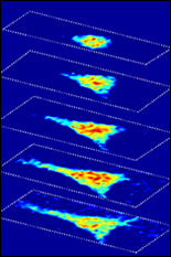
To target a probe beam of light beyond the diffraction limit, Scarcelli and BIOE postdoctoral researcher Eitan Edrei essentially wrapped a second beam of light of a different wavelength around the probe. This allowed the team to create transient microlenses with a diameter equal to the optical diffraction limit. As a result, when the probe beam passes through the sculpted microlens, it zeroes in on a tighter focal point than otherwise allowed by the optical system. In this way, the probe of light focuses beyond the system’s diffraction barrier.
 “We demonstrated a potential way to increase the focusing ability of an optical system,” Scarcelli said. “Even more, we have shown the ability to transiently manipulate the property of a material using a second beam of light, which could open up many possibilities for tunable and flexible optical systems."
Scarcelli’s work represents the first experimental demonstration of focusing and imaging beyond the diffraction limit of a given optical system – without the need to add to or alter the observed sample itself. Because of this, their method is broadly compatible with imaging and focusing systems used in many applications, such as in photochemistry or in optical trapping – a process whereby scientists use a highly focused laser beam to grasp and move microscopic objects as if they were using a tiny pair of tweezers.
“We are now working on creating a new paradigm for ‘lenses’ in which the optical performance is decoupled from the geometrical form,” Scarcelli said. “For example, this means that we could impart desired optical functions to elements that are conformable to irregularly-shaped surfaces – which could point to revolutionary advancements in integrated photonics and point-of-care sensing.”
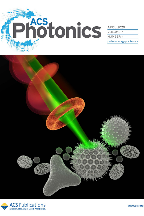
Scarcelli’s work was featured this week in Optics in 2020, a special issue of the Optical Society of America dedicated to highlight the best optics research of the year. The full study was first published in an ACS Photonics paper titled “Optical Focusing beyond the Diffraction Limit via Vortex-Assisted Transient Microlenses” earlier this year. Endrei served as first author on the ACS Photonics paper.
Scarcelli is also a faculty member with the Robert E. Fischell Institute for Biomedical Devices.
Published December 3, 2020
Related Stories
Stories / June 29, 2017
New Microscopy Technique Sheds Light on How Cells Sense...
Stories / October 5, 2015
Novel Microscopy Technique to Shed New Light on Study of Cell...
Stories / March 4, 2022
Olympus Discovery Center Inaugurated at UMD

Stories / August 16, 2021
Using Light to Attack Cancer

Stories / June 15, 2020
Scarcelli Applies NSF CAREER Award to Study Embryonic...
Stories / August 26, 2019
Scarcelli Receives Junior Faculty Outstanding Research Award
Stories / March 11, 2019
New Microscopy Technique Could Change LASIK

Stories / September 21, 2018
New pyroelectric system transmits power wirelessly, harvesting...
Stories / April 23, 2018
Three BIOE Professors Awarded NIH R01s in as Many Months
Stories / March 1, 2018
BIOE Postdoc Recognized for Early Colon Cancer Detection...